Personal collections

Explain how X-rays are produced for use in medical diagnosis.
State why X-ray images are taken of multiple sections of the body during computed tomography (CT) scanning.
An X-ray image is taken of the structure shown in the figure below.
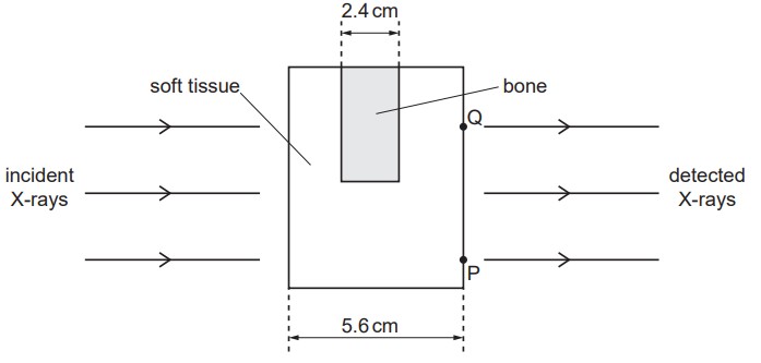
The linear attenuation coefficient of bone is  . The linear attenuation coefficient of soft tissue is
  . The incident X-rays are parallel and have a uniform intensity
  across the structure. Determine, in terms of , the intensity of the detected X-rays from:
point P
point Q.
Explain, with reference to your answers in (b), whether the X-ray image of the structure in the figure above has good contrast.


 .
. .
.