Personal collections

Electrons are accelerated through a potential difference of  . They are then incident on a metal target, they decelerate, and X-ray photons are emitted.
Calculate the maximum possible frequency of the emitted X-ray photons.
Explain why an aluminium filter may be placed in the X-ray beam when producing an X-ray image of a patient.
The linear attenuation (absorption) coefficients  for X-rays in bone, blood, and muscle are given in the table below.
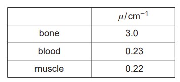
A beam of these X-rays is incident on a person. Calculate the percentage of the intensity of the X-ray beam that has been absorbed after passing through  of blood.
In an X-ray image, white regions show greater absorption of X-rays than dark regions. State and explain the difference between the X-ray image of bone compared to that of muscle.


 .
. .
.